Anatomy III: The muscles
Muscles give the body its surface forms. Each one has an origin (the fixed end), an insertion (the end that moves) and an action. When a muscle contracts it gets shorter and thicker; when it stretches it flattens. That is what you draw.
How muscles change shape
A flexed biceps bunches into a high ball; a straight arm shows a long, flat form. Muscles on one side of a joint contract while their opposites relax and stretch — so a bent elbow shows a bulging front and a stretched back.
Tendons are flat and cord-like. Where a muscle turns into a tendon the surface form changes from soft and rounded to hard and linear — think of the back of the knee or the front of the wrist.
Groups rather than names
Learn muscles in groups that make one shape: the shoulder (deltoid), the chest (pectoralis), the belly wall (rectus abdominis and obliques), the thigh front (quadriceps) and the calf (gastrocnemius and soleus). Each group has a clear silhouette you can hang details on.
Draw shapes that wrap around the forms beneath. A muscle never sits flat on the surface; it follows the cylinder of the limb or the curve of the ribcage.
Reference table
| Muscle | Location | Function | What you see |
|---|---|---|---|
| Sternocleidomastoid | Neck, behind ear to breastbone | Turns and tilts the head | Two diagonal cords forming a V at the pit of the neck |
| Trapezius | Upper back and neck | Lifts and pulls the shoulders back | The slope from neck to shoulder |
| Deltoid | Shoulder cap | Raises the arm | Rounded shoulder; a dimple where it inserts on the arm |
| Pectoralis major | Chest | Pulls the arm across the body | Fan shape that forms the front of the armpit |
| Latissimus dorsi | Side and lower back | Pulls the arm down and back | The V-shape of the back |
| Biceps brachii | Front of upper arm | Bends the elbow, turns the palm up | Bulges when the arm flexes |
| Triceps brachii | Back of upper arm | Straightens the elbow | Horseshoe shape above the elbow |
| Forearm flexors / extensors | Forearm | Bend and straighten the wrist and fingers | Two long masses that twist as the hand turns |
| Rectus abdominis | Front of abdomen | Bends the trunk forward | Segmented 'six-pack' between the ribs and pubis |
| External oblique | Sides of abdomen | Twists and bends the trunk | Soft bulge above the hip bone |
| Gluteus maximus | Buttock | Extends the hip | The main mass of the buttock |
| Quadriceps | Front of thigh | Straightens the knee | Teardrop of vastus medialis above the inner knee |
| Sartorius | Diagonal across the thigh | Crosses the legs | A long strap dividing front and inner thigh |
| Hamstrings | Back of thigh | Bend the knee | Tendons visible either side of the back of the knee |
| Gastrocnemius | Calf | Points the foot | Two heads; inner one lower than outer |
| Tibialis anterior | Front of shin | Lifts the foot | Soft form just outside the sharp shin bone |
Historic plates
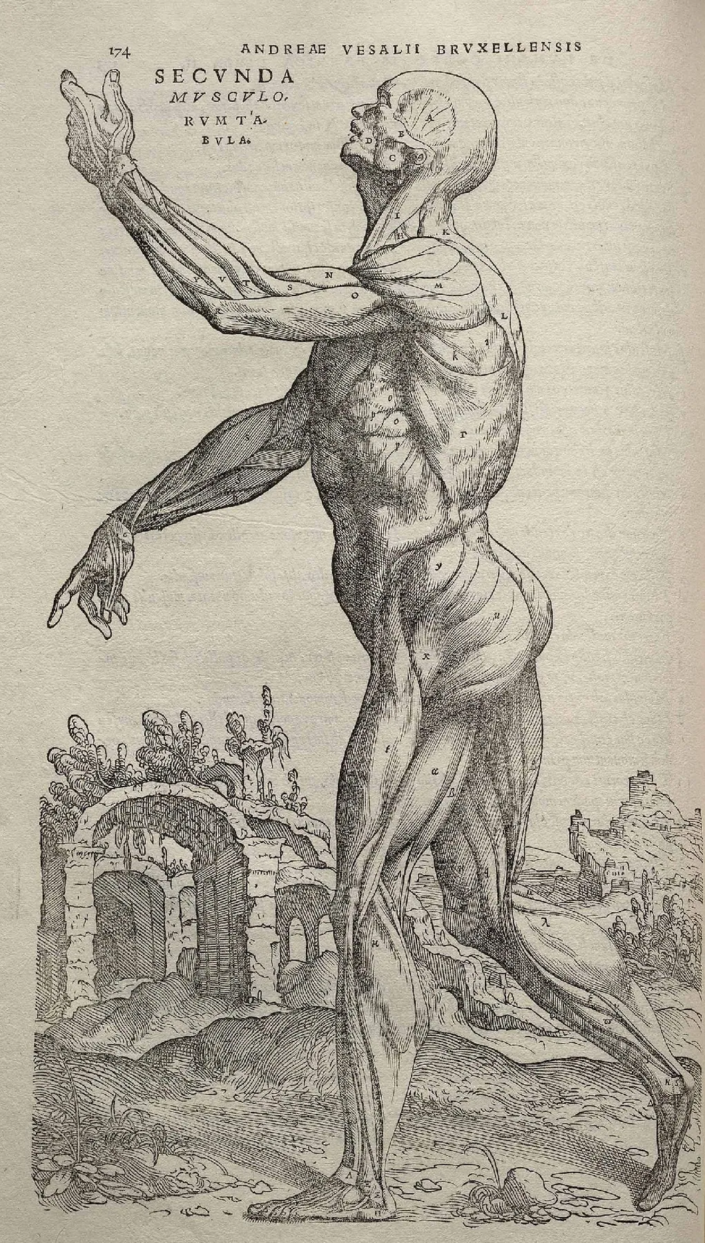
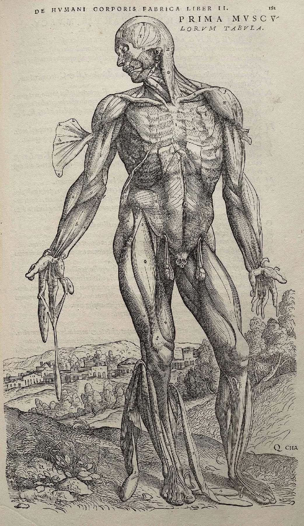
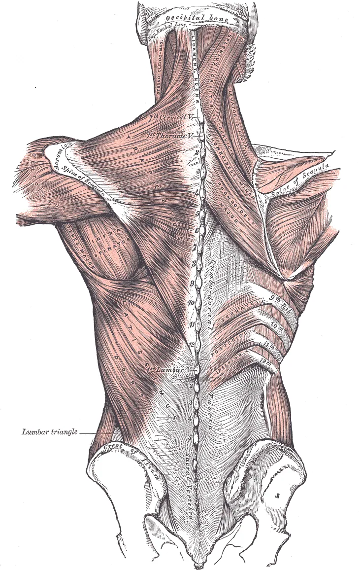
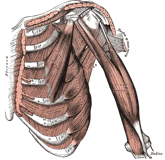
Public-domain plates from Vesalius (1543), Leonardo's anatomical notebooks and Gray's Anatomy (1918). Tap to zoom into the detail.
Master studies
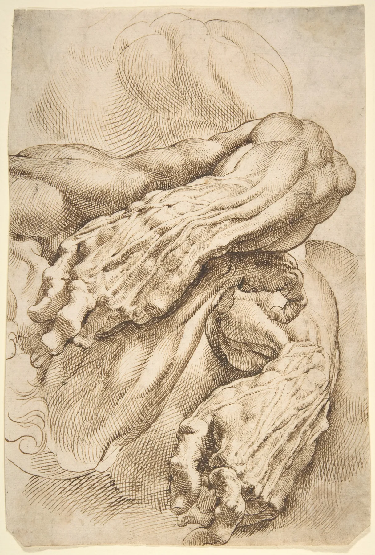
- Draw your own arm bent and straight from a mirror and compare the biceps and triceps shapes.
- Trace a photo of an athlete and outline only the major muscle groups from the table.
Watch
How to Draw Pecs - Anatomy · Proko
How to Draw the Upper Back Muscles - Anatomy and Motion · Proko
How to Draw the Neck - Anatomy for Artists · Proko
Videos load from YouTube (privacy-enhanced mode) only when you press play.