Home / Anatomy

Anatomy for artists

Skeleton, muscles, head and skull, torso, hands, feet, movement and foreshortening — with names, locations and functions, clear diagrams and public-domain plates by Vesalius, Leonardo and Gray.

Idealised 8-head figure proportion with landmarks at each head unit12345678ChinNipplesNavel / elbowsCrotch / wrists (mid-point)Mid-thigh / fingertipsBelow kneesMid-shinSoles of feet≈ 2 heads wide

Proportion and the figure

Artists measure the body in head-lengths. Real people vary — most adults are about 7 to 7½ heads tall — but the idealised 8-head figure give…

Labelled schematic of the human skeleton, front viewCraniumMandible (jaw)ClavicleSternumHumerusPelvis (ilium)RadiusCarpals + handFemurPatellaTibiaCervical spineRibcage (12 pairs)Lumbar spineUlnaSacrum / pubisHip jointFibulaTarsals + footSchematic front view · dashed lines = head units (8 heads tall)

The skeleton

The skeleton is the scaffold that sets proportions and shows through the skin at key points. An adult has about 206 bones, but an artist nee…

Simplified surface muscles of the body, front view, labelledSternocleidomastoidTrapeziusPectoralis majorBiceps brachiiRectus abdominisForearm flexorsAdductorsRectus femorisVastus medialisTibialis anteriorDeltoidExternal obliqueSartoriusVastus lateralisGastrocnemius (calf)Simplified surface muscles, front view (shapes stylised for artists)

The muscles

Muscles give the body its surface forms. Each one has an origin (the fixed end), an insertion (the end that moves) and an action. When a mus…

Labelled schematic of the skull, side viewFrontal boneBrow ridgeOrbit (eye socket)Nasal boneMaxillaMandibleChin (mental protuberance)Parietal boneOccipital boneTemporal boneZygomatic archMastoid processAngle of jawSkull, side view (schematic). Dashed: sutures between plates

The head and skull

Every portrait sits on a skull. Learn the few planes and landmarks of the skull and you can draw a head from any angle — then add the featur…

Ribcage and pelvis as two boxes, neutral and in contrappostoRibcagePelvisNeutralRibcagePelvisContrappostoTwo rigid masses joined by the flexible spine (blue). In contrappostothey tilt in opposite directions.

The torso

The torso is two boxes — ribcage and pelvis — linked by a flexible column. Almost every expressive pose comes from tilting, twisting or bend…

Bones of the hand, schematic, labelledCarpals (8 wrist bones)Thumb metacarpalMetacarpals (palm)Thumb: 2 phalangesPhalanges (3 per finger)Knuckle arc (dashed)WristHand bones, back view (schematic). 27 bones; the knuckles form an arc,not a straight line.

Arms and hands

Hands are as expressive as faces and just as feared. Treat the hand as a box-shaped palm with a wedge-shaped thumb and four tapering fingers…

Bones of the foot from the inner side, schematic, labelledTibiaTalus (ankle)Calcaneus (heel)FibulaNavicular + cuneiformsMetatarsalsInner arch (dashed)Phalanges (toes)Foot, inner side (schematic). Weight rests on the heel and the ball; theinner arch lifts between them.

Legs and feet

Legs carry the weight and feet meet the ground. Get these right and a figure stands; get them wrong and it floats.…

Foreshortening: cylinders at three angles and overlapping formsSide onTiltedPointing at youOverlapnear → farAs a limb points toward you it gets shorter and its end ellipses getrounder. Near forms overlap far ones.

Movement and foreshortening

Movement is anatomy in action. Foreshortening is what happens when a form points towards or away from you — and it is the hardest and most r…

All anatomical plates

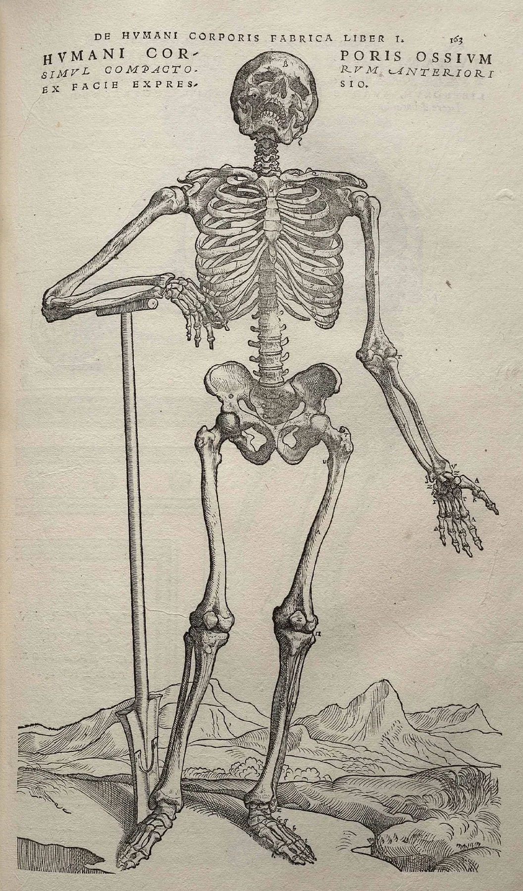
Skeleton, front view (De humani corporis fabrica) by Andreas Vesalius (woodcut attributed to the workshop of Titian)
Skeleton, front view (De humani corporis fabrica), Andreas Vesalius (woodcut attributed to the workshop of Titian) (1543). Plate / drawing. Source: Wikimedia Commons · Public domain
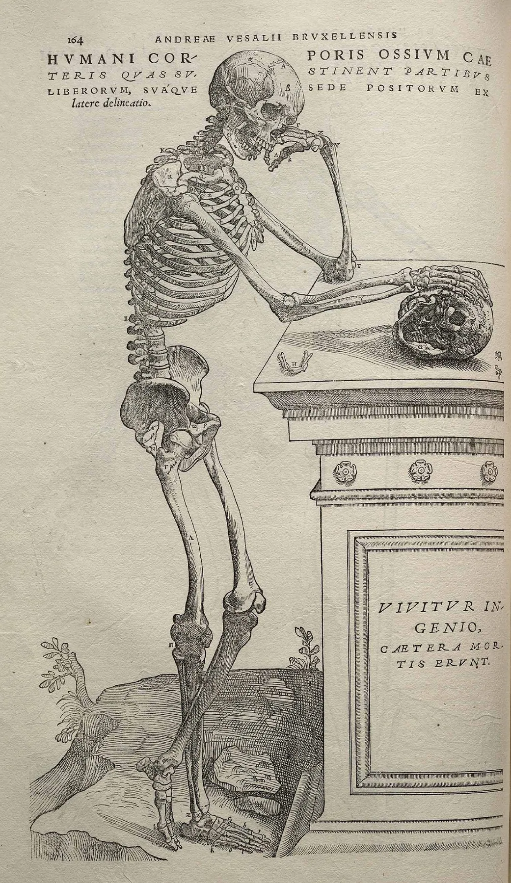
Skeleton, side view (De humani corporis fabrica) by Andreas Vesalius
Skeleton, side view (De humani corporis fabrica), Andreas Vesalius (1543). Plate / drawing. Source: Wikimedia Commons · Public domain
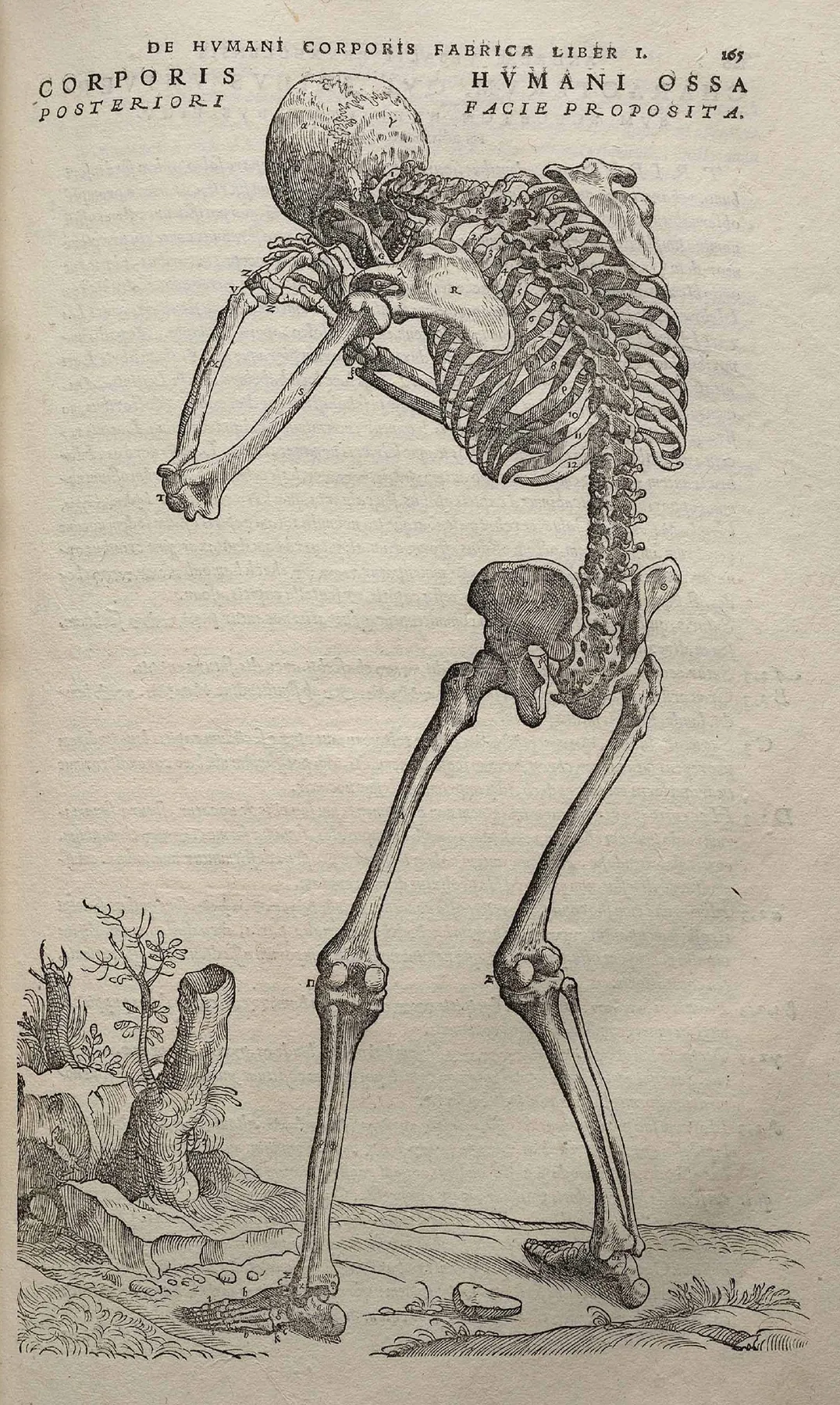
Skeleton, back view (De humani corporis fabrica) by Andreas Vesalius
Skeleton, back view (De humani corporis fabrica), Andreas Vesalius (1543). Plate / drawing. Source: Wikimedia Commons · Public domain
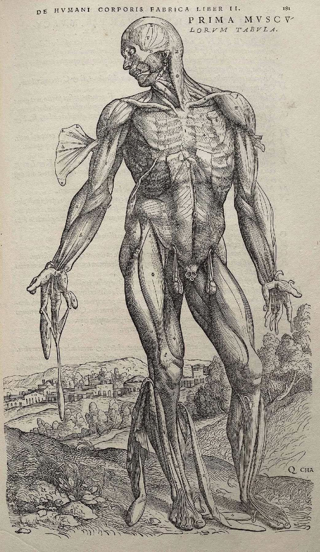
First plate of the muscles (De humani corporis fabrica) by Andreas Vesalius
First plate of the muscles (De humani corporis fabrica), Andreas Vesalius (1543). Plate / drawing. Source: Wikimedia Commons · Public domain
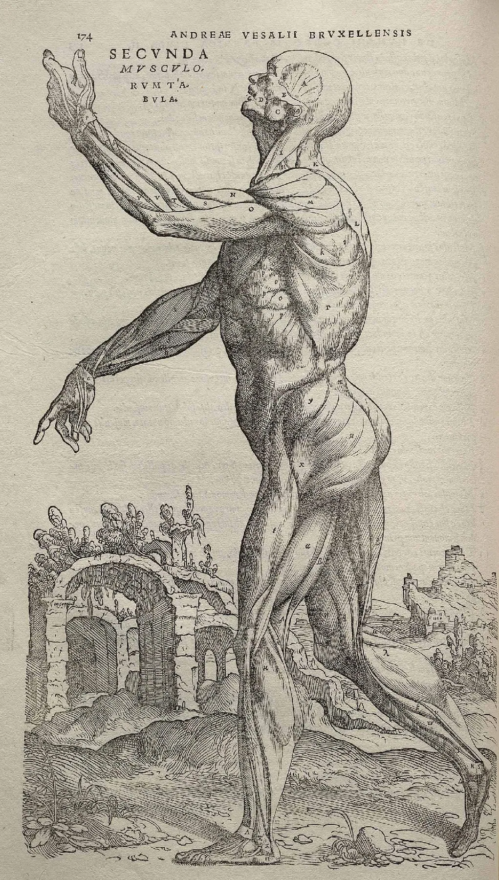
Muscle plate (De humani corporis fabrica) by Andreas Vesalius
Muscle plate (De humani corporis fabrica), Andreas Vesalius (1543). Plate / drawing. Source: Wikimedia Commons · Public domain
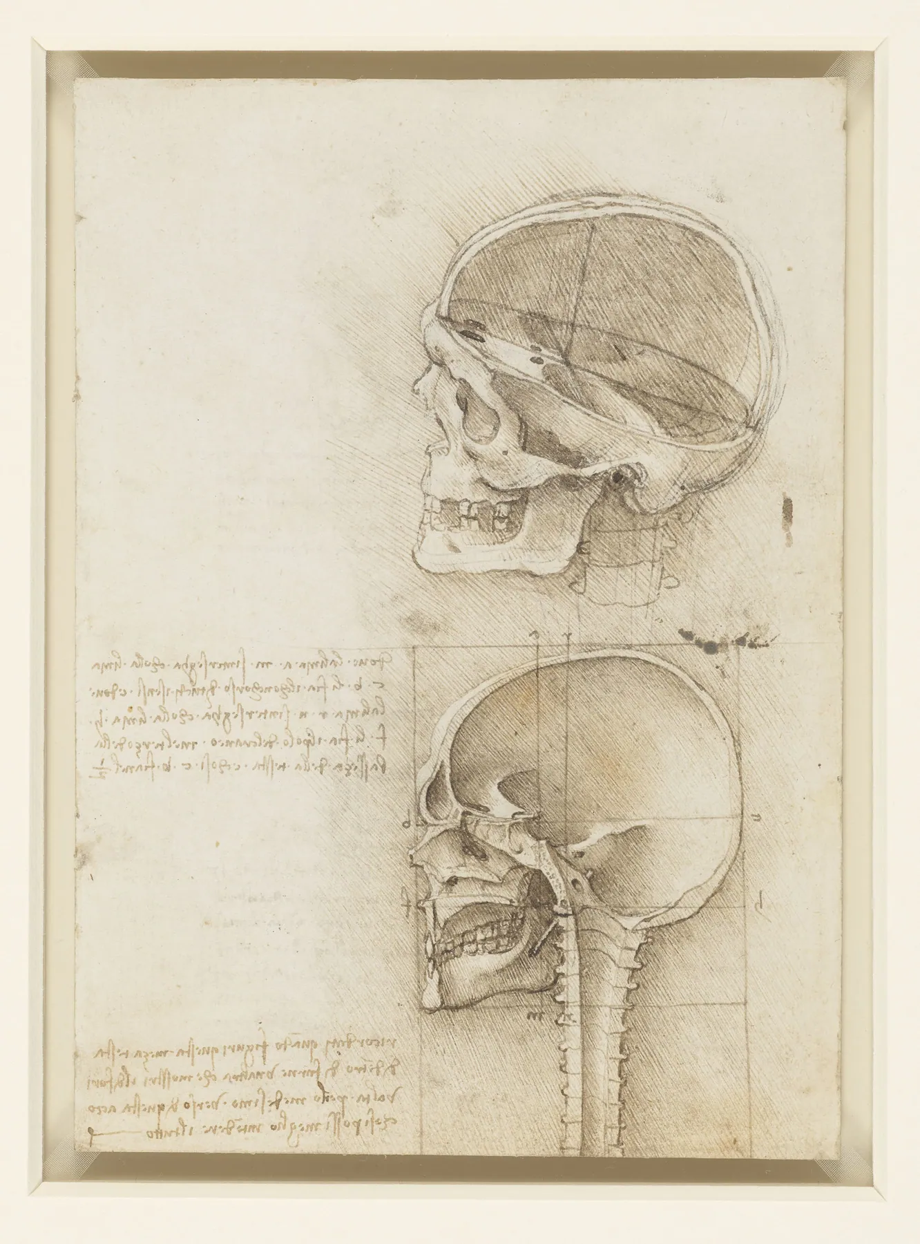
The skull sectioned by Leonardo da Vinci
The skull sectioned, Leonardo da Vinci (1489). Plate / drawing. Source: Wikimedia Commons · Public domain
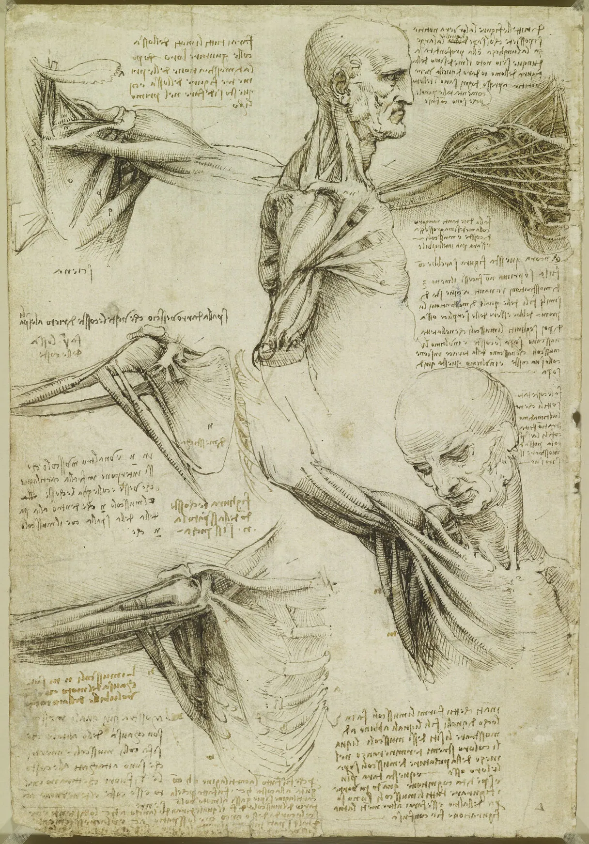
The muscles of the shoulder by Leonardo da Vinci
The muscles of the shoulder, Leonardo da Vinci (c. 1510–1511). Plate / drawing. Source: Wikimedia Commons · Public domain
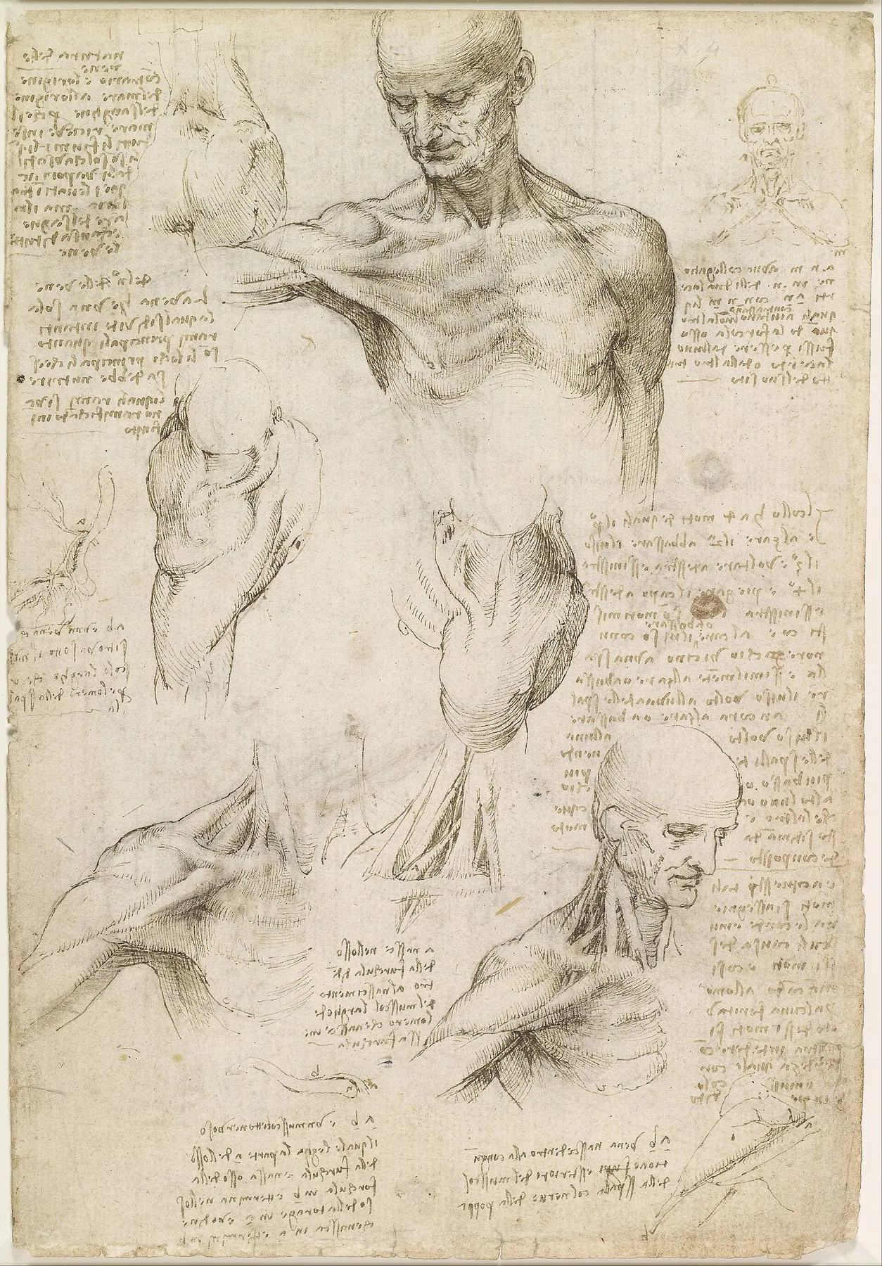
Superficial anatomy of the shoulder and neck by Leonardo da Vinci
Superficial anatomy of the shoulder and neck, Leonardo da Vinci (c. 1510–1511). Plate / drawing. Source: Wikimedia Commons · Public domain
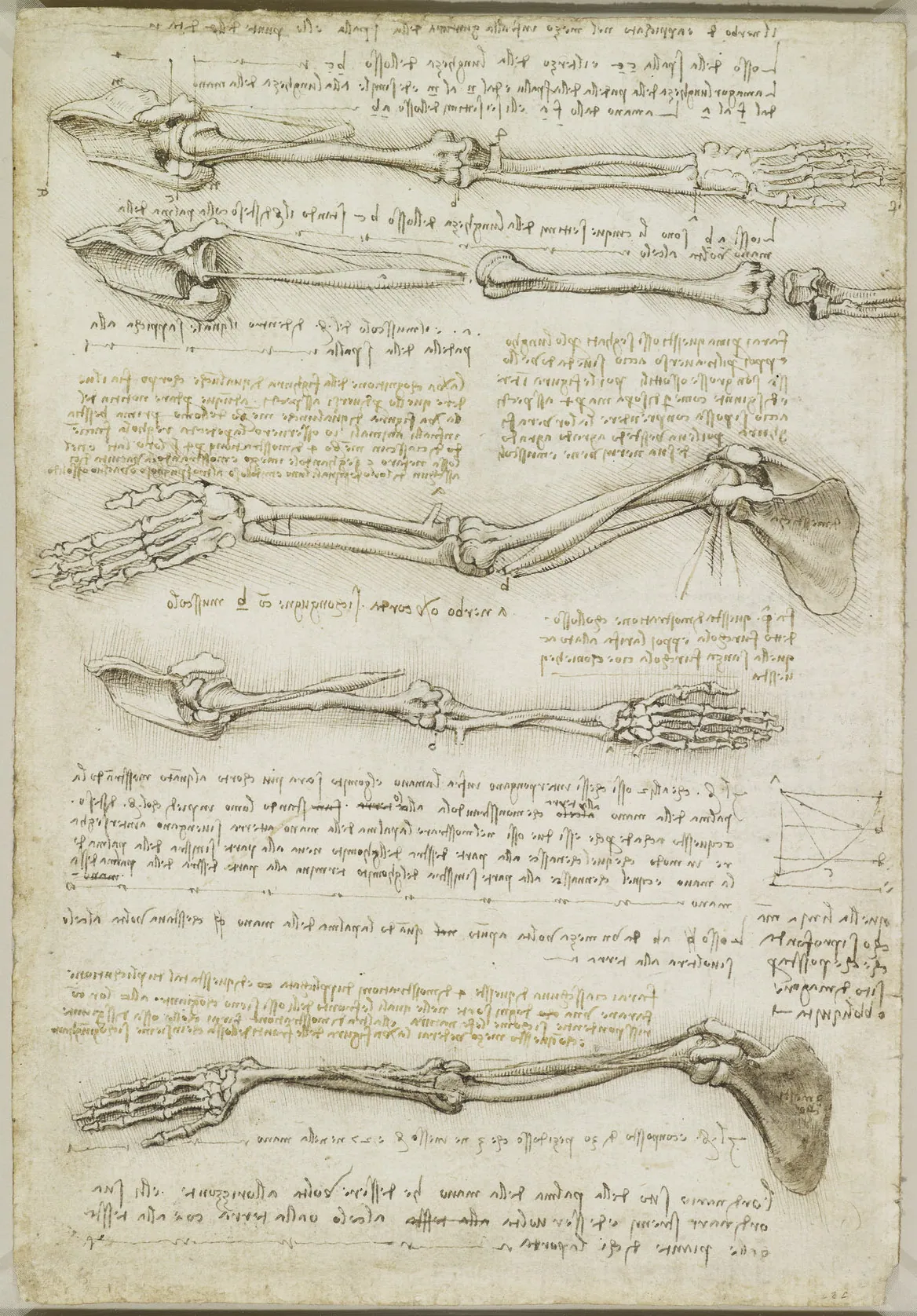
The bones and muscles of the arm by Leonardo da Vinci
The bones and muscles of the arm, Leonardo da Vinci (c. 1510–1511). Plate / drawing. Source: Wikimedia Commons · Public domain
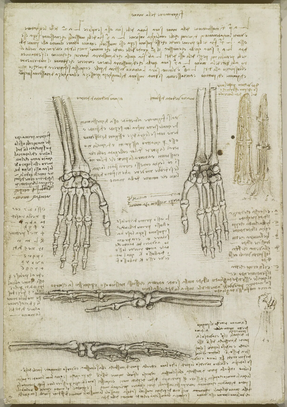
The bones of the hand by Leonardo da Vinci
The bones of the hand, Leonardo da Vinci (c. 1510–1511). Plate / drawing. Source: Wikimedia Commons · Public domain
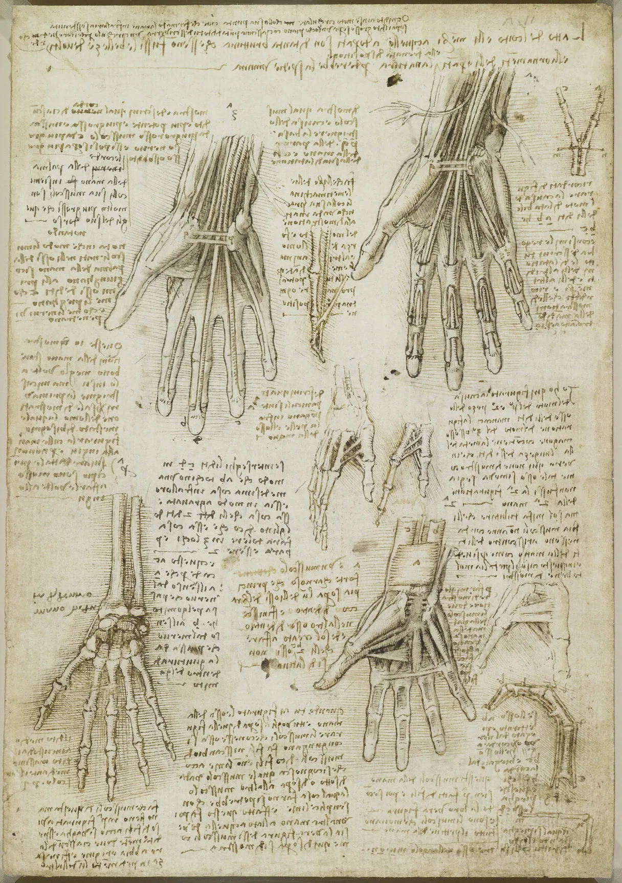
The bones, muscles and tendons of the hand by Leonardo da Vinci
The bones, muscles and tendons of the hand, Leonardo da Vinci (c. 1510–1511). Plate / drawing. Source: Wikimedia Commons · Public domain
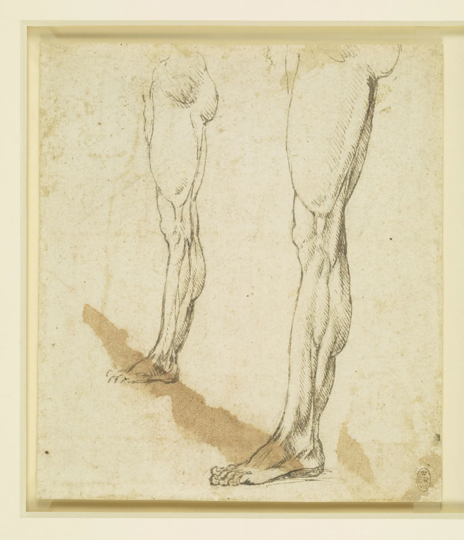
Two studies of a man's left leg in profile by Leonardo da Vinci
Two studies of a man's left leg in profile, Leonardo da Vinci (c. 1500s). Plate / drawing. Source: Wikimedia Commons · Public domain
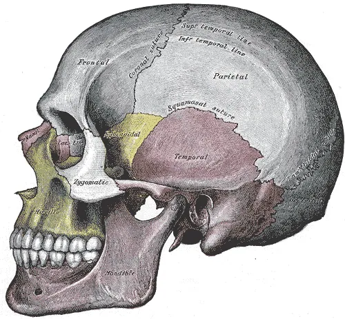
Skull, side view (Gray's Anatomy, fig. 188) by Henry Vandyke Carter for Henry Gray
Skull, side view (Gray's Anatomy, fig. 188), Henry Vandyke Carter for Henry Gray (1918 edition). Plate / drawing. Source: Wikimedia Commons · Public domain
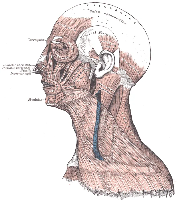
Muscles of the head, face and neck (Gray's Anatomy, fig. 378) by Henry Vandyke Carter for Henry Gray
Muscles of the head, face and neck (Gray's Anatomy, fig. 378), Henry Vandyke Carter for Henry Gray (1918 edition). Plate / drawing. Source: Wikimedia Commons · Public domain
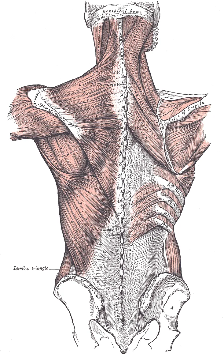
Muscles of the back (Gray's Anatomy, fig. 409) by Henry Vandyke Carter for Henry Gray
Muscles of the back (Gray's Anatomy, fig. 409), Henry Vandyke Carter for Henry Gray (1918 edition). Plate / drawing. Source: Wikimedia Commons · Public domain
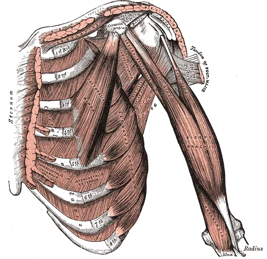
Superficial muscles of the chest and arm (Gray's Anatomy, fig. 411) by Henry Vandyke Carter for Henry Gray
Superficial muscles of the chest and arm (Gray's Anatomy, fig. 411), Henry Vandyke Carter for Henry Gray (1918 edition). Plate / drawing. Source: Wikimedia Commons · Public domain
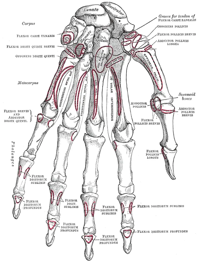
Bones of the left hand, palm view (Gray's Anatomy, fig. 219) by Henry Vandyke Carter for Henry Gray
Bones of the left hand, palm view (Gray's Anatomy, fig. 219), Henry Vandyke Carter for Henry Gray (1918 edition). Plate / drawing. Source: Wikimedia Commons · Public domain
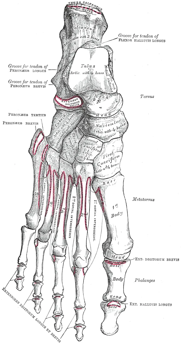
Bones of the left foot, top view (Gray's Anatomy, fig. 268) by Henry Vandyke Carter for Henry Gray
Bones of the left foot, top view (Gray's Anatomy, fig. 268), Henry Vandyke Carter for Henry Gray (1918 edition). Plate / drawing. Source: Wikimedia Commons · Public domain
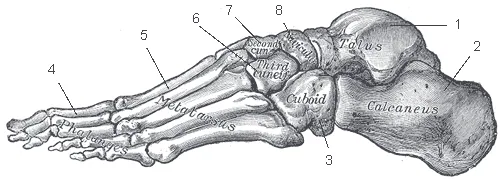
Bones of the foot, outer side (Gray's Anatomy) by Henry Vandyke Carter for Henry Gray
Bones of the foot, outer side (Gray's Anatomy), Henry Vandyke Carter for Henry Gray (1918 edition). Plate / drawing. Source: Wikimedia Commons · Public domain

Practise

Proportion-spotting game · Proportion guide tool · Gesture drill timer