Anatomy VII: Legs and feet
Legs carry the weight and feet meet the ground. Get these right and a figure stands; get them wrong and it floats.
The leg
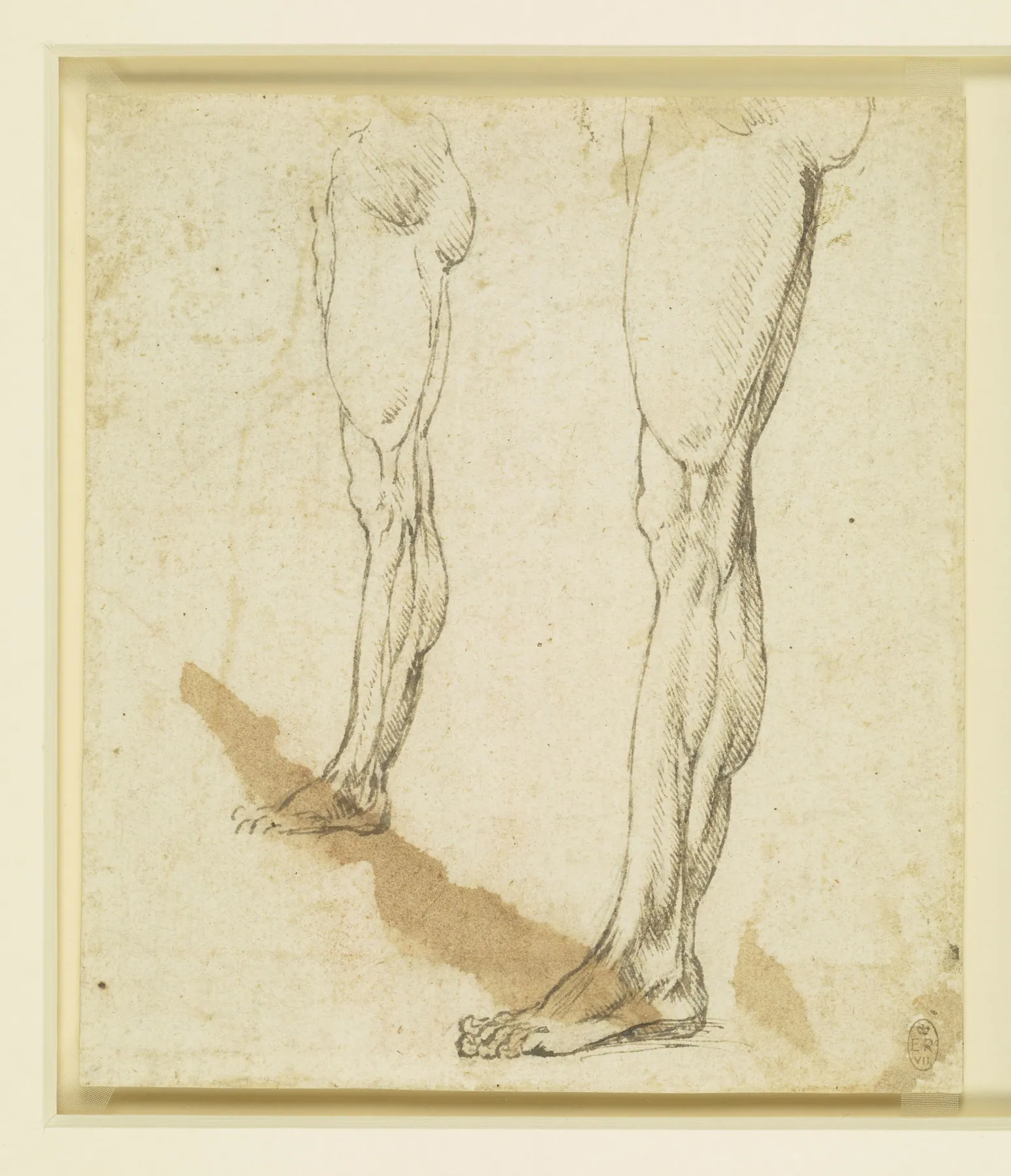
The thigh bone angles inward from hip to knee, so the knees sit closer together than the hips. The outer thigh curves more than the inner one high up; the inner calf is lower and fuller than the outer. These alternating curves give the leg its rhythm.
The knee is a box with the kneecap set into it. From the side the leg is not straight: the thigh curves forward, the shin is straight at the front and the calf curves behind.
The foot
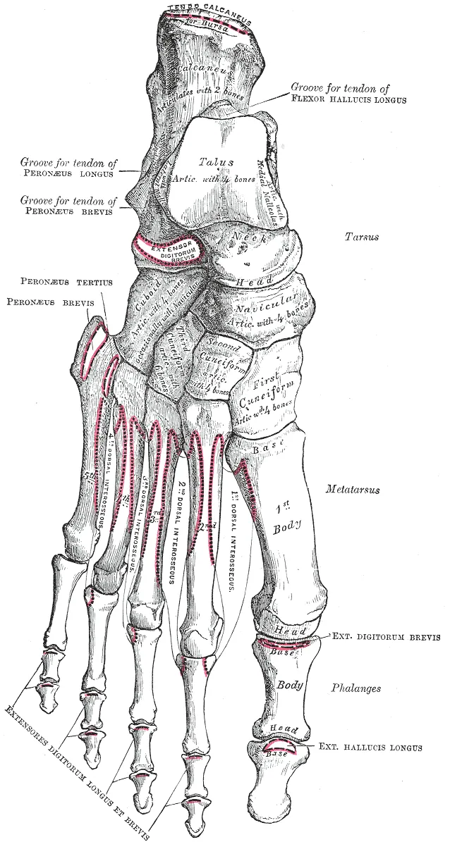
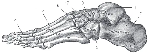
The foot is a wedge: high at the ankle, low at the toes. The inner side has a high arch; the outer edge stays nearly flat on the ground. The inner ankle bone sits higher than the outer one.
Weight rests on the heel and the ball of the foot. Draw the foot first as a simple block, then cut the toes into the front at an angle — the big toe is longer and points straight ahead, the others curl down.
Reference table
| Part | Bones / muscles | Note |
|---|---|---|
| Thigh | Femur; quadriceps, hamstrings, adductors | Angles inward toward the knee |
| Knee | Patella, femur, tibia | Square block from the front |
| Lower leg | Tibia, fibula; calf and shin muscles | Sharp shin, full calf |
| Ankle | Talus; malleoli | Inner bump high, outer bump low |
| Heel | Calcaneus | Carries the weight at the back |
| Arch | Tarsals and metatarsals | High inside, low outside |
| Toes | Phalanges | Big toe has 2 bones, the others 3 |
Historic plates
Public-domain plates from Vesalius (1543), Leonardo's anatomical notebooks and Gray's Anatomy (1918). Tap to zoom into the detail.
Master studies
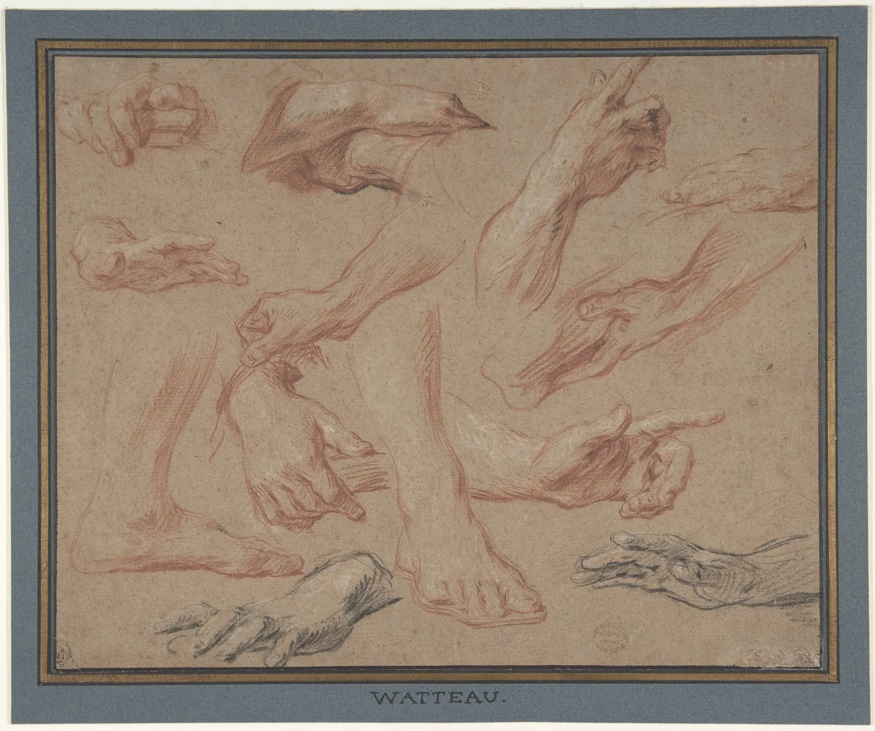
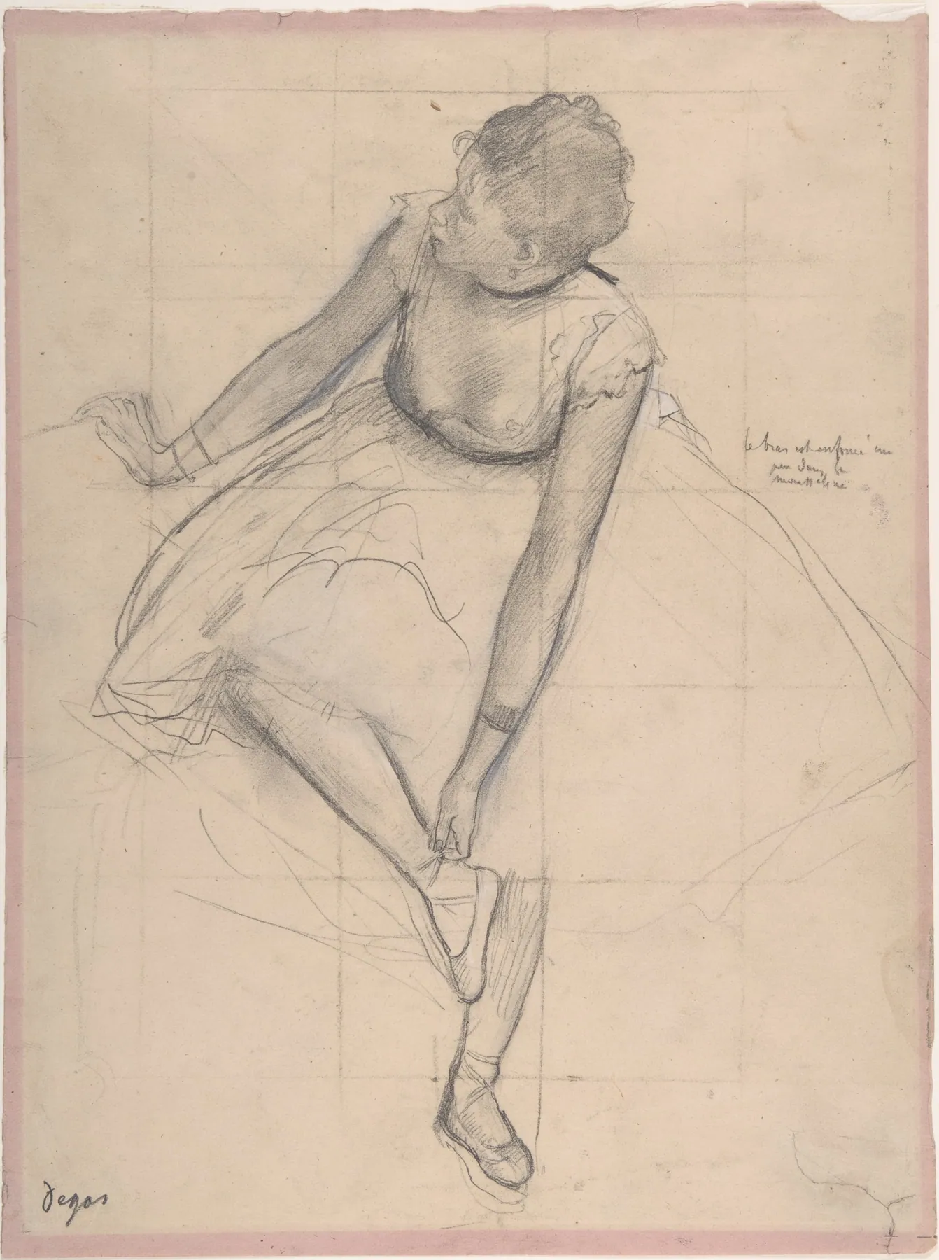
- Draw your own foot from the side and from the front, as a block first.
- Copy the Leonardo leg studies, tracing the alternating curves.
Watch
How to Draw Feet with Structure - Foot Bone Anatomy · Proko
How to Draw Feet · Proko
Simplifying the Foot for Easy Drawing · Proko
Videos load from YouTube (privacy-enhanced mode) only when you press play.